17 July 2000
Hajime Kawakami
Life
time performance of FM-intensifier in analog mode
1.
Introduction
The previous
report (Swift-UVOT/MSSL.TC/0002) showed capability of analog mode for high
time resolution photometry, when a target star was brighter than 17.4mag.
Swift UVOT may have several bright stars in the field of view during monitoring
time variation of a gamma ray burster. It is dangerous for the intensifier
to observe a bright star for a long time in photon counting mode (high
gain in MCPs). The analog mode observation will offer the longer life time,
since the electric gain of MCPs is less than 1/6 of the photon counting
mode.
Quantifying
very bright star as a stand alone system is one of the safety requirements
for Swift UVOT. Analog mode is one of the candidate to fulfil this requirement.
It is, however, essential to know, how long does the detector withstand
against the intense star illumination during the brightness assessment.
Our intensified
CCD detector demonstrated far longer life time (>100 times) than typical
position sensitive detectors in terms of accumulated anode charge (XMM-OM/MSSL/TC/
0059). This difference may be due to the lower gain of MCPs with our detector,
i.e. ~5x10^5 with ours while ~10^7 with the position sensitive detectors.
If the life time depends on the gain strongly, the reduction of the gain
by the factor of 6 may extend the life time far longer than x6.
In this
report, the image intensifier was operated in the analog mode and was exposed
to intense pinhole illuminations for 100 hours. Gain depletion of MCPs
and sensitivity loss in F-F images were investigated against accumulated
charge.
2.
Electric gain
The best
result in analog mode was obtained with 90% of nominal photon counting
MCP voltage in the test with DEP_#5 intensifier. The experiments in this
report was carried out with DEP_#8 intensifier, whose nominal MCPs voltage
is 2250V. The 2020V, 90% of the nominal, was applied to the MCPs during
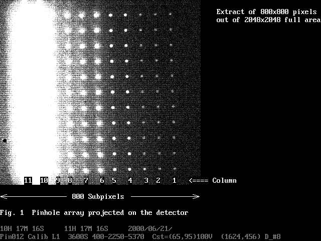
the photon dose. The illumination pattern was a 11x11 pinhole array and
the brightness has gradient along the column in the dynamic range of ~2E+4
(Fig. 1). The light source is made of 64 green LEDs covered with a diffuser
and a 5300-5700A interference filter. The brightness of the LEDs is controlled
by a constant current source in the dynamic range of ~2E+4 (see detail
in XMM-OM/MSSL.TC/0057).
The
brightness of the pinhole array was calibrated with3 photon counting images
in the 3 LED current levels, L=1, L=3 and L=10 in order to overcome small
dynamic range of the detector. The lower LED current (L=1) was used for
determining brightness ratio among bright pinhole columns, while the medium
LED current (L=3) was for faint pinhole columns (Table 1). The highest
current (L=10) was for the calibration of LED brightness during the photon
dose, in which the faintest pinhole column (col=1) still gave useful data.
Photon losses due to coincidence were corrected and the true input rates
were tabulated in Table 2. Finally, the absolute brightness of the pinhole
columns at the LED current level of L=10 was tabulated in Table 3.
Table 1.
Raw counts /(hour x spot) 21 June 2000 DEP_#8
-------------------------------------
LED =
1
3
10
-------------------------------------
col=11
264920.0
N/A
N/A
col=10
254720.0
N/A
N/A
col=
9 32620.0
N/A
N/A
col=
8 24460.0
N/A
N/A
col=
7
2381.2
N/A
N/A
col=
6
2131.9 23104.0
N/A
col=
5
158.2
1680.4
N/A
col= 4
84.1
1042.4
N/A
col=
3
22.5
186.2
N/A
col=
2
( 7.0)
169.0
(253160.0)
col=
1
14.2
164.6
264110.0
-------------------------------------
Table
2. True counts /(sec x spot) 21 June 2000 DEP_#8
-------------------------------------
LED =
1
3
10
-------------------------------------
col=11
94.58122
col=10
90.11536
col=9
9.74329
col=8
7.26678
col=7
.69737
col=6
.62426
6.85783
col=5
.04627
.49191
col=4
.02459
.30502
col=3
.00658
.05446
col=2
(.00205)
.04942 (89.43997)
col=1
.00415
.04814
94.22338
-------------------------------------
Table
3. Pinhole brightness at LED current level = 10
---------------------------------------------------------------
column
1 2
3 4
5 6
7 8
9 10
11
---------------------------------------------------------------
Brtness
94.22 96.73 106.59 597.0
962.8 13.4k 15.0k 156k
209k 1938k 2034k
(c/s)
B0 star
16.5 16.5 16.4
14.5 14.0 11.1
11.0 8.4
8.1 5.7
5.6
(mag)
---------------------------------------------------------------
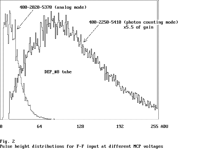
The ratio
of gains between V_mcp=2250V(photon counting) and 2020V(analog) were determined
from both of the brightness of event splash at phosphor screen and anode
current. The pulse height distributions of the event splash with the 2
different voltages to MCPs were shown in Fig. 2. Flat filed in the count
rate of 15,000 c/(sec full area) were used for the illumination. The brief
ratio was determined from the peak positions, > 5.5 times. It was difficult
to determine the ratio accurately, because the pulse height distribution
with V_mcp=2020V was squashed to the lower energy end.
The brighter
F-F illumination was used for the measurement of the anode current to produce
sufficient current with V_mcp=2020V. The detected count rate for the F-F
was 86,100 c/(sec full area) in photon counting mode. After the correction
of the coincidence loss, the true incoming rate was turned out to be 94,000
c/(sec full area). The procedure of the coincidence correction followed
XMM-OM/MSSL.TC/0050. Coincidence area of event splashes was assumed to
be 12 (CCD_pixels)^2 from other 2 intensifiers, though there was no data
on DEP_#8 itself. The full detector area in the photon counting imaging
is (3.37 x 256 CCD_pixels)^2, while photocathode area is circle with the
diameter of 25mm. Since the anode current was produced from all photocathode
area, incoming rate of electrons involved in anode current was 94,000 c/s
* (D=25mm) / (3.37 x256 (CCD_pixels)^2 = 94,000 c/s * 1.2467 = 117,000
c/s. A 99.91k Ohm resister was inserted at the anode cable, whose voltage
was at 8000V, and the small voltage drop across the resister was measured
with a precision multimeter, FLUKE 87 IV, in the minimum readout of 1uV.
The resistance value was also calibrated by the FLUKE 87 IV. The small
voltage drops were 1012uV and 151uV with V_mcp=2250V and 2020V. Hence,
the currents were 10.23nA and 1.53nA. Since the input impedance of Fluke
87IV was 10M Ohm, anode currents were corrected by the factor of 1.01.
Finally, the electric gain was calculated to be 5.4x10E+5 with V_mcp=2250V
and 8.1x10E+4 with V_mcp=2020V. The ratio of the gains with the 2 voltages
was x6.7 times.
The electric
gain in the high input rate was measured using pinhole illuminations in
the LED current levels of L=1-10 for V_mcp=2250V and L=3-10 for V_mcp=2020V.
Columns=1-9 of the pinhole array was blocked for this measurement, so that
the brightest 2 columns=10-11 with nearly same brightness were used for
the illumination. Since voltage display of the FLUKE 87 IV was not stable
in the last 2 digits (10uV, 1uV), the display was read 10 times and was
averaged for the lowest 2 illuminations (i.e. LED current levels L=3 and
L=4 for V_mcp=2020V and L=1 and L=2 for V_mcp=2250V). The results for the
both of V_mcp=2250V and V_mcp=2020V were tabulated in Table 4 and were
plotted in Figs. 3 and 4. The electric gain of the intensifier was 5.7E+5
in the low input rate with V_mcp = 2250V and 8.1E+4 with V_mcp = 2020V.
The gain depletion is 1/9.7 in the input rate of 2E+6 c/s with V_mcp =
2250V, while 1/8 with V_mcp = 2020V, compared with those at the input rate
of 100 c/s. The electric gains of pinhole illumination in the low input
rate was higher than that of F-F illumination. This was due to global gain
variation of MCPs ( i.e. the local gain at pinhole positions were higher
than the average).
Electric
gain of MCPs at pinhole positions should have changed during the photon
dose. The anode current was measured after completing the 100 hours photon
dose by illuminating exactly same positions. This gauges the level of the
change before and after the photon dose. Again, columns=1-9 of the pinhole
array was blocked for the measurement, so that the brightest 2x11 pinholes
from columns=10-11 were used for the illumination. The gains at the brightest
pinhole positions at different input rate were tabulated in Table 5 and
were plotted in Figs. 3 and 4 overlaying on the original gains. In spite
of the large gain loss in the low input rate, the gain in the high input
rate does not change before and after the 100 hours dose. This is particularly
true for the illumination above 1E+5 c/(sec x spot) with V_mcp = 2250V
and 1E+6 c/(sec x spot) with V_mcp = 2020V. From these results, we can
assume anode currents at columns=10 and 11 were constant throughout the
photon dose, hence we can estimate total accumulated charge precisely.
There is no measurement on the change of gain at other pinhole positions,
i.e. columns=1-9. Since the total accumulated charges are smaller, the
anode currents hopefully did not change much before and after the photon
dose.
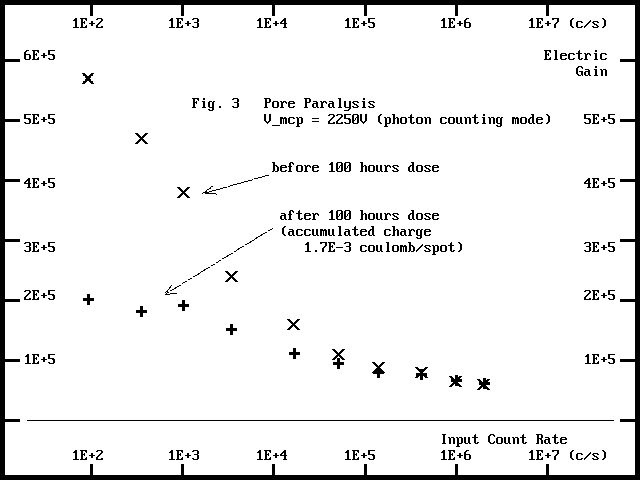
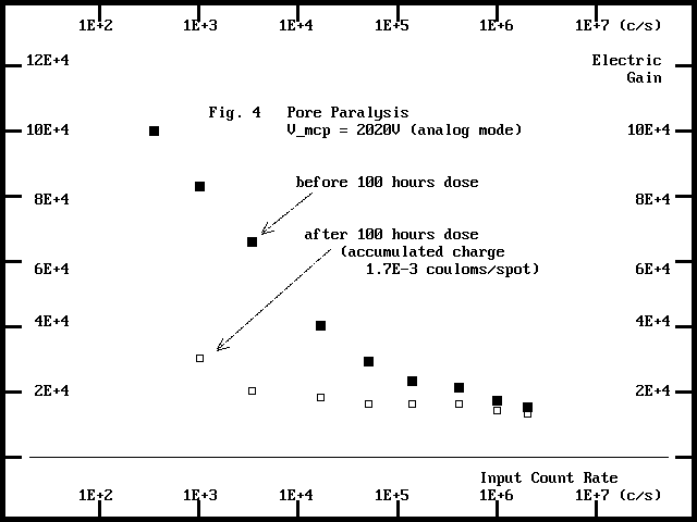
Because
of the large gain loss in the low input rate while no gain loss in the
high input rate after the 100 hours photon dose, the gradient of the gain
curve against input rate becomes flat. This suggests a very hard scrubbing
may lighten the effect of pore paralysis, hence may extend dynamic range
of MCPs.
Table 4.
Electric gain of XMM-OM tube in high count rate
--------------------------------------------------------
LED Intensity Anode
current (pA)
Electric Gain
(c/s pinhole)
from 22 pinholes
2020V 2250V
2020V 2250V
--------------------------------------------------------
F-F
94000
1530 10230
8.1 E+4 5.4 E+5
L=1
92.35
(6.7)
184
(2. E+4) 5.7 E+5
L=2
352
132
585
10. E+4 4.7 E+5
L=3
1014
295
1370
8.3 E+4 3.8 E+5
L=4
3426
800
2880
6.6 E+4 2.4 E+5
L=5
16500
2330
9260
4.0 E+4 1.6 E+5
L=6
51000
5250 20200
2.9 E+4 1.1 E+5
L=7
139000
11100 43300
2.3 E+4 0.88E+5
L=8
410000
29700 114000
2.1 E+4 0.79E+5
L=9
984000
57800 225000
1.7 E+4 0.65E+5
L=10
1986000
102000 412000
1.5 E+4 0.59E+5
--------------------------------------------------------
Table 5. Electric gain after 100 hours
dose
--------------------------------------------------------
LED Intensity Anode
current (pA)
Electric Gain
(c/s pinhole)
from 22 pinholes
2020V 2250V
2020V 2250V
--------------------------------------------------------
L=1
92.35
---
64
---
2.0 E+5
L=2
352
---
219
---
1.8 E+5
L=3
1014
103
664
3. E+4 1.9
E+5
L=4
3426
239
1810
2. E+4 1.5
E+5
L=5
16500
1041
6550
1.8 E+4 1.1
E+5
L=6
51000
2790 16900
1.6 E+4 0.94E+5
L=7
139000
7700 38400
1.6 E+4 0.78E+5
L=8
410000
24000 108000
1.6 E+4 0.75E+5
L=9
984000
50200 225000
1.4 E+4 0.65E+5
L=10
1986000
94100 419000
1.3 E+4 0.60E+5
-------------------------------------------------------------------
Ref-2
Files used in this section
/swift/ZPHD010.dat
ZPIN011.dat,ZPIN012.dat,ZPIN013.dat,ZPIN014.dat
3.
Gain loss of MCPs
A reference
pulse height distributions for individual pinhole columns, col=4-11 (600-2E+6
c/s), were measured with V_mcp=2250V (photon counting mode) before starting
the photon dose. The photon doses were carried out 3 times for 15 hours,
15 hours and 70 hours with V_mcp=2020V. The pulse height distributions
were measured after the each photon dose. Fig. 5 shows the reference pulse
height distribution and the one after the 100 hours dose by the 2E+6 c/s
pinholes.
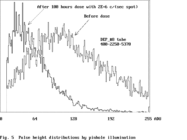
The gain
reduced to 1/2.5 of the beginning. The gain loss was quantified from peak
positions of the pulse height distributions. The gains after each dose
were tabulated in Table 6, and were plotted against accumulated charge
in Fig 6. Fig. 7 is the extract from XMM-OM/MSSL.TC/ 0059, in which the
intensifier was operated in photon counting mode during the photon dose.
The plots in analog mode coincides with those in photon counting mode.
This implies that the gain loss can be described by the single parameter,
accumulated
charge, in any condition (i.e. different gain, input count rate, exposure
time etc.).
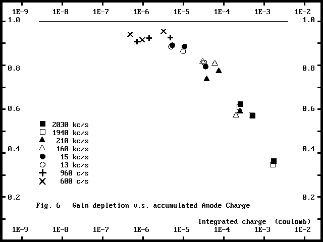
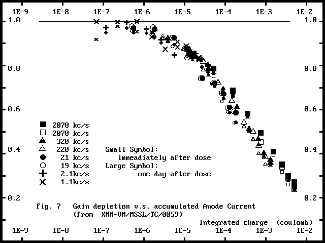
Table 6.
Gain depletion of MCPs
----------------------------------------------------------
Dose
Pinhole intensity (counts/sec)
time
2030k 1940k
210k 160k
15k 13k
960 600
----------------------------------------------------------
15.0 hr
.623 .609
.735 .816
.890 .884
.907 .941
30.0 hr
.570 .573
.772 .807
.884 .862
.922 .916
100.0
hr .363
.346 .589
.570 .793
.811 .924
.955
----------------------------------------------------------
Ref-3 Files
used in this section
/swift/ZPHD016.dat,ZPHD028.dat,ZPHD047.dat,ZPHD064.dat
4.
Sensitivity loss in photon counting image
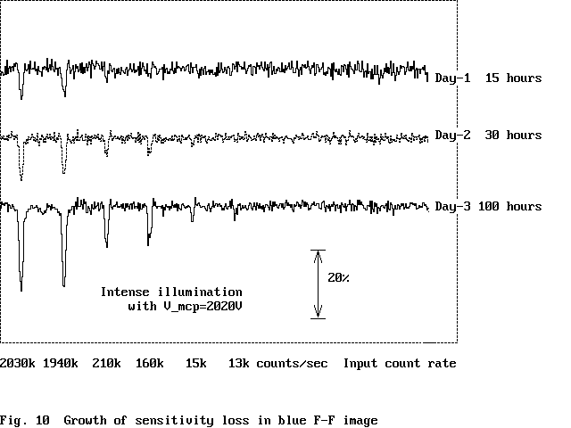
A reference
F-F image with the blue (460nm) LED illumination was integrated for 15
hours in photon counting mode before starting the photon dose. F-F image
integrations were followed after each intense illumination to see the sensitivity
losses in the different level of dose. The integrations started after the
sufficient period since the end of the intense illumination in order to
avoid fluorescence, i.e. 80 hours in the 1st day, 38 hours the 2nd day
and 27 hours the last day.
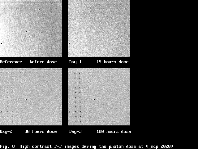
Fig. 8 shows
4 F-F images, one was taken prior to the photon dose for reference and
the other 3 were after 15, 30 and 100 hours dose. The F-F after 100 hours
dose clearly showed an array of black spots corresponding to the pinhole
positions.
A F-F image
in each day of photon dose was divided by the reference F-F to remove detector
artefacts and illumination non-uniformity. Then, the 11x11 array of black
spots were averaged along the columns to improve S/N. Central positions
of the black spots coincided with the pinhole positions in the accuracy
of 8um along H_intensifier direction (bias direction of the 1st MCP), while
systematically shifted by 15um along V_intensifier direction. The day by
day growth of the black spots is shown in Fig. 9. The image contains all
factors, i.e. fluorescence, gain depletion and photocathode sensitivity
loss. Fig. 10 shows profiles of the averaged black spots in the last day.
Y-width of the slice is 3 twixel (= 58um). The depth of black spots reached
25% at the brightest pinhole after 100 hours dose. The black spots were
noticeable for the illumination intensities of > 13kc/s after 100 hours
dose, but not for the lower intensity illuminations. Since the F-F integrations
were started after the sufficiently long period since the end of the photon
dose, the peak depths were only little affected by fluorescence (less than
1.4%).
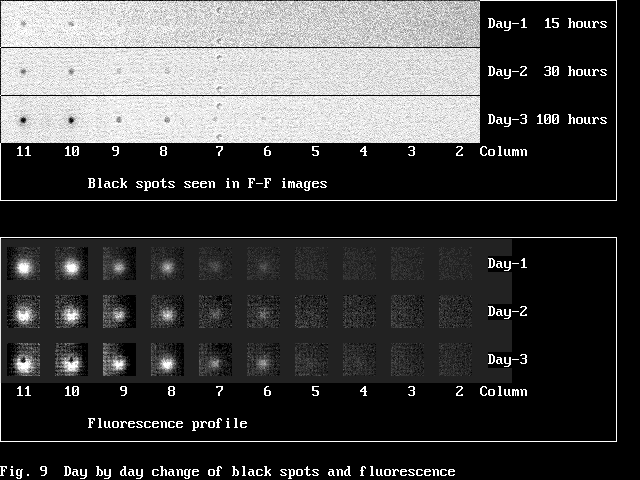
The 6-10
dark frames were acquired in photon counting mode before and after the
F-F integrations in order to correct the effect of fluorescence for further
precision. Standard fluorescence profiles are shown in the lower panel
of Fig. 9. The size of the fluorescence is far larger than both of the
black spots and projected pinhole, i.e. from 220um to 270um. The fluorescence
spots have offset from the pinhole positions by 200-240um along H_intensifier
direction (bias direction of the 1st MCP). The fluorescence spot got doughnut
shape (i.e. black spot in central part) with the increase of the dose level.
The details of these characteristics were identical to those described
in the former report, XMM-OM/MSSL/TC/0057.
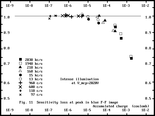
The sensitivity
loss at the peak position was quantified from the average of 3x3 twixels
square centred on the black spots. The normalization level was determined
from 37x37 twixels (=717um) square excluding central D=21 twixels circular
area. Then, the effect of fluorescence (1.4 in maximum) was subtracted.
The results were tabulated in Table 7 and were plotted against accumulated
charge in Fig. 11. The sensitivity did not decrease till 1E-5 couloms/spot.
It started to decrease steeply from 1E-4 couloms/spot.
The sensitivity
loss seen in F-F image is the combination of gain depletion and photocathode
sensitivity loss. The photocathode sensitivity losses were calculated by
removing the effect of gain depletion (threshold=15ADU). The results were
tabulated in Table 8 and were shown in Fig. 12. The sensitivity loss of
photocathode is not noticeable up to 3E-5 couloms/spot.
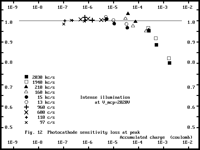
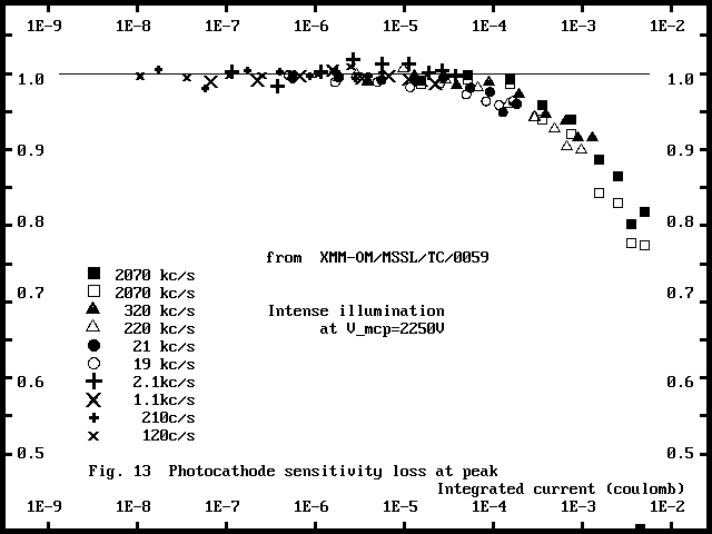
The photocathode
sensitivity loss for the same intensifier but dosed in photon counting
mode was extracted from XMM-OM/MSSL/TC/0057 (Fig. 13). The sensitivity
loss dosed in analog mode shows slightly faster decrease above 2E-4 couloms/spot.
This result did not imply that ion barrier characteristics of the 1st MCP
improved with the lower voltage.
A Gaussian
profile was fitted to the black spots to quantify the spatial extent. The
results are shown in Fig. 14. There are clear correlation with accumulated
charge. The width increased from 80um(FWHM) to 110um after acquiring 1E-5
to 2E-3 couloms/spot.
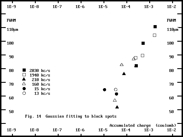
Table 7.
Sensitivity change in blue F-F image at peak position
----------------------------------------------------------------------
Dose
Pinhole intensity (counts/sec)
time
2030k 1940k
210k 160k
15k 13k
960 600
110 97
----------------------------------------------------------------------
15.0 hr
.933 .942
1.000 .993
1.002 1.010
1.006 1.001
.999 .984
30.0 hr
.864 .892
.971 .958
.979 .995
.994 1.002
1.000 1.001
100.0
hr .743
.755 .907
.910 .956
.977 .997
.999 1.001
.997
-----------------------------------------------------------------------------------------------------
Table 8.
Photocathode sensitivity change at peak position
----------------------------------------------------------------------
Dose
Pinhole intensity (counts/sec)
time
2030k 1940k
210k 160k
15k 13k
960 600
110 97
----------------------------------------------------------------------
15.0 hr
.951 .965
1.032 1.009
1.007 1.016
1.014 1.006
.999 .984
30.0 hr
.886 .917
.994 .975
.985 1.001
1.001 1.009
1.000 1.001
100.0
hr .801
.823 .955
.950 .966
.983 1.004
1.003 1.001
.997
----------------------------------------------------------------------
Ref-4
Files used in this section
/swift/ZDEP015.dat,ZDEP030.dat,ZDEP043.dat,ZDEP060.dat
ZPHD016.dat,ZPHD028.dat,ZPHD047.dat,ZPHD064.dat
ZDRK019.dat,ZDRK020.dat,ZDRK021.dat,ZDRK029.dat,ZDRK031.dat
ZDRK032.dat
ZDRK033.dat,ZDRK034.dat,ZDRK035.dat,ZPHD036.dat,ZPHD037.dat
ZDRK038.dat,ZDRK039.dat,ZDRK042.dat,ZPHD044.dat,ZPHD045.dat
ZDRK048.dat,ZDRK049.dat,ZDRK050.dat,ZPHD051.dat,ZPHD052.dat
ZDRK053.dat,ZDRK054.dat,ZDRK055.dat,ZPHD059.dat,ZPHD061.dat
5.
Summary
i) Gain
loss dosed in analog mode was same as that in photon counting mode in terms
of accumulated anode charge.
ii) Photocathode
sensitivity loss dosed in analog mode was slightly faster (by ~30%) than
that in photon counting mode in terms of accumulated anode charge.
iii) Gain
loss measured in the low input rate was 1/2 - 1/3 after 100 hours photon
dose, while that in the high input rate almost nothing. In the consequence,
pore paralysis curve was flatten after the dose. This suggests a possibility
of extending dynamic range of MCPs by a hard scrubbing.
Appendix.
Experiment procedure for DEP_#8 intensifier in analog mode
20 June - 6 July 2000
-------------------------------------------------------------------
File Name
Pinhole
PHD
Dark
F-F
Time(start)
-------------------------------------------------------------------
Before Damage for reference
2000/06/20
PHD010
300FRs
17H 51M 05S
Pin011 L=3
54000S
19H 01M 34S
Pin012
L=1
3600S
10H 17M 16S
Pin013
L=10
1800S
15H 11M 25S
Pin014
L=10
1800S
16H 21M 36S
DEP015
54000S
18H 13M 44S
PHD016
70000FRs
11H 27M 18S
Ana017
10000FRs
12H 37M 00S
Ana018
10000FRs
16H 50M 13S
\/\/\/\/\/\/\\/\/\/\/\/\/\/\/\/\/\/\/\/\/\/\/\/\/\/\/\/\/\/\/\/\/\/\/\/\/\/\/\/\/\\/\/\/\/\/\/\/\/\/\/\/\/\/\/\/\/\/\/\/\/\/\/\/\/\/\/\/\/\/
15 hour
Day-1
18:24 - 09:24
2000/06/22
\/\/\/\/\/\/\//\/\/\/\/\/\/\/\/\/\/\/\/\/\/\/\/\/\/\/\\/\/\/\/\/\/\\/\/\/\/\/\/\/\/\/\/\/\/\/\/\/\/\/\/\/\/\/\/\/\/\/\/\/\//\/\/\/\/\/\/\/\/
2000/06/23
Drk019
7200S
10H 36M 54S
Drk020
7200S
12H 37M 18S
Drk021
7200S
14H 37M 42S
DEP022
Th=15 54000S
17H 48M 35S
Drk023
7200S
08H 48M 59S
Drk024
7200S
10H 49M 22S
Drk025
7200S
12H 49M 45S
Drk026
7200S
14H 50M 08S
Ana027
20000FRs
12H 28M 17S
PHD028
70000FRs
14H 34M 34S
Drk029
7200S
15H 37M 46S
DEP030
Th=15 54000S
17H 38M 10S
2000/06/27
Drk031
7200S
08H 38M 34S
Drk032
7200S
10H 38M 58S
\/\/\/\/\/\/\\/\/\/\/\/\/\/\/\/\/\/\/\/\/\/\/\/\/\/\/\/\/\/\/\/\/\/\/\/\/\/\/\/\/\\/\/\/\/\/\/\/\/\/\/\/\/\/\/\/\/\/\/\/\/\/\/\/\/\/\/\/\/\/
15 hour
Day-2
13:15 - 04:16
2000/06/27
\/\/\/\/\/\/\//\/\/\/\/\/\/\/\/\/\/\/\/\/\/\/\/\/\/\/\\/\/\/\/\/\/\\/\/\/\/\/\/\/\/\/\/\/\/\/\/\/\/\/\/\/\/\/\/\/\/\/\/\/\//\/\/\/\/\/\/\/\/
2000/06/28
Drk033
7200S
12H 41M 57S
Drk034
7200S
19H 37M 23S
Drk035
7200S
21H 37M 47S
Drk036
7200S
23H 38M 11S
Drk037
7200S
01H 38M 35S
Drk038
7200S
03H 38M 59S
Drk039
7200S
05H 39M 23S
PHD040
50000FRs
10H 41M 54S
-------------------------------------------------------------------
File Name
Pinhole
PHD
Dark
F-F
Time(start)
-------------------------------------------------------------------
Ana041
30000FRs
11H 23M 58S
Drk042
7200S
16H 52M 29S
DEP043
54000S
18H 52M 53S
Drk044
7200S
09H 53M 17S
Drk045
7200S
11H 53M 40S
Drk046
7200S
13H 54M 03S
PHD047
50000FRs
17H 24M 09S
\/\/\/\/\/\/\\/\/\/\/\/\/\/\/\/\/\/\/\/\/\/\/\/\/\/\/\/\/\/\/\/\/\/\/\/\/\/\/\/\/\\/\/\/\/\/\/\/\/\/\/\/\/\/\/\/\/\/\/\/\/\/\/\/\/\/\/\/\/\/
70 hour
Day-3
18:05 - 16:05
2000/06/30
\/\/\/\/\/\/\//\/\/\/\/\/\/\/\/\/\/\/\/\/\/\/\/\/\/\/\\/\/\/\/\/\/\\/\/\/\/\/\/\/\/\/\/\/\/\/\/\/\/\/\/\/\/\/\/\/\/\/\/\/\//\/\/\/\/\/\/\/\/
Drk048
7200S
17H 20M 36S
Drk049
7200S
19H 21M 01S
Drk050
7200S
21H 21M 25S
Drk051
7200S
23H 21M 49S
Drk052
7200S
01H 22M 13S
Drk053
7200S
03H 22M 37S
Drk054
7200S
05H 23M 01S
Drk055
7200S
07H 23M 25S
Drk056
7200S
09H 23M 49S
Drk057
7200S
11H 24M 13S
Drk058
7200S
13H 24M 37S
Drk059
7200S
15H 25M 01S
DEP060
54000S
19H 26M 24S
Drk061
7200S
10H 26M 48S
Drk062
7200S
12H 27M 12S
Ana063
30000FRs
15H 47M 40S
PHD064
50000FRs
09H 39M 36S
-------------------------------------------------------------------------------